As in nerve conduction studies, a need exists in
needle examination to develop broad-range work-ups
designed for general groups of pathological processes.
Along with that, a working knowledge of the spinal
segments of the upper and lower extremities' muscles
is an absolute prerequisite for adequate interpretation
of the needle examination.
A broad range work-up allows:
- A nonbiased approach to the patient's problem leaving
open the possibility that a disease other than the
referral diagnosis may be found.
A simplified approach to general groups of diseases
that can be tailored to fit the particular process
at hand.
- Five general work-ups which are in general, similar
but not identical to those described in the section
on nerve conduction studies are thus described. They
are: routine upper extremity, routine lower extremity,
peripheral neuropathy, anterior horn cell disease,
and myopathy.
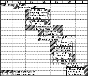
Routine Upper Extremity
Designed for the study of roots, plexus, entrapment,
and traumatic neuropathies of the upper extremity, this
work-up emphasizes sampling of muscles belonging to
different upper extremity nerves and innervated by root
levels C5-T1.
The work-up consists of sampling the following (or
other similarly innervated) muscles:
- The first dorsal interosseous (an ulnar C8, T1 muscle)
- The flexor pollicis longus (an anterior interosseous
C7,8 muscle)
- The flexor carpi radialis (a median C7 muscle)
- The brachioradialis (a radial C5,6 muscle)
- The triceps (a radial C7,8 muscle)
- The deltoid (an axillary C5,6 muscle).
In the root lesions work-up, the appropriate paraspinal
levels should be sampled.
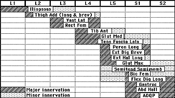
Routine Lower Extremity
Designed for the study of roots, plexus, entrapment,
and traumatic neuropathies of the lower extremity this
work-up emphasizes sampling of muscles belonging to
different lower extremity nerves and innervated by root
levels L3-S2.
The work-up consists of sampling the following (or
other similarly innervated) muscles:
- The extensor digitorum brevis or extensor hallucis
longus (peroneal L5-S1 muscles)
- The flexor digitorum longus (a posterior tibial
L5-S1,2 muscle)
- The tibialis anterior (a peroneal L4,5 muscle)
- The medial gastrocnemius (a posterior tibial S1,2
muscle)
- The vastus lateralis (a femoral L3,4 muscle)
- The gluteus medius (a superior gluteal L4,5 and
S1 muscle)
In the root lesions work-up, the appropriate paraspinal
levels should be sampled.
Peripheral Neuropathy
This work-up, which emphasizes distal muscles sampling
because these are usually more involved in the typical
neuropathic processes, consists of:
- A routine upper extremity examination with an extra
distal muscle included, the abductor digiti minimi
- A routine lower extremity examination with the abductor
hallucis included
Anterior Horn Cell
The main goal of this work-up is to sample muscles from
a widespread root distribution to rule out the possibility
of multiple motor radiculopathies. A minimum of two
routine extremities work-ups need to be done.
These should include:
- A routine upper extremity
- A routine lower extremity
- A third upper or lower extremity depending on the
areas of clinical involvement
- The tongue
Myopathy
For the study of the different groups of myopathies
including the myotonias and the Lambert-Eaton syndrome,
this work-up consists of modified routine upper and
lower extremities studies with an emphasis on proximal
muscles.
This should include:
- A modified routine upper extremity with the flexor
pollicis longus deleted and the biceps and infraspinatus
added
- A modified routine lower extremity with the flexor
digitorum longus deleted and thigh abductors and iliacus
added.
- In the inflammatory myopathies, the paraspinal muscles
are usually quite involved and their sampling increases
the diagnostic yield.
Neuromuscular Transmission
Single fiber EMG has greatly altered the traditional
neuromuscular transmission defects work-ups by needle
electrodes. Through moment to moment variation in the
shape and amplitude of affected motor unit potentials
is helpful, jitter analysis by single fiber EMG is a
much more sensitive means to study defects in neuromuscular
transmission. The technique requires the use of a special
needle electrode which has a 25 µm tip on a side
port to allow recording from single muscle fibers. When
the tip is positioned in the vicinity of two muscle
fibers belonging to the same motor unit, two potentials
are seen firing synchronously. If, by means of a delay
line and a signal trigger, one of them is made to trigger
the sweep, the distance between the two potentials,
or interpotential interval, is observed to vary from
discharge to discharge. The distance between the first
and second potential is measured over a certain number
of tracings and the mean interpotential interval is
calculated. The standard deviation around that mean
or the mean of the consecutive differences (MCD) are
used in expressing the jitter which to a large extent
represents the variability in neuromuscular transmission.
In neuromuscular transmission disorders, the jitter
is increased early in the course of the illness, before
repetitive stimulation tests become positive. In the
later stages, impulse blocking due to total failure
in neuromuscular transmission is seen and results in
the disappearance of one of the potentials on the screen.