|
|
|
|
Motor
Responses
The motor response is obtained
by stimulating a nerve and recording from a muscle that
it innervates. The muscle selected should have a fairly
well-defined motor point, and preferably be relatively
isolated from other muscles innervated by the nerve and
from other nerves that may be stimulated inadvertently
during the test. The excitation of nearby muscles may
alter the response and make it difficult to determine
the exact onset of the desired motor response.
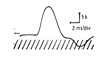 The
motor response may be characterized by its amplitude,
duration, and wave form. The amplitude is measured from
the baseline to the top of the negative peak of the
motor response and is expressed in millivolts. The
motor response may be characterized by its amplitude,
duration, and wave form. The amplitude is measured from
the baseline to the top of the negative peak of the
motor response and is expressed in millivolts.
The distal latency is measured
from the onset of the stimulus artifact to the point
of takeoff from the baseline and is measured in milliseconds.
Extra care must be taken to use the corresponding takeoff
points of both the distal and proximal responses so
that conduction velocities are measured along the same
fibers. The amplitude depends to a large extent on the
number and size of muscle fibers being activated, and
supramaximal stimulation of the nerve should ensure
a maximal motor response. Any pathological process that
decreases the number of motor units or muscle fibers
responding will affect the amplitude. The normal motor
response indicates a fairly synchronous discharge of
the motor units. If there is dispersion of the times
when the motor units discharge, then the amplitude will
be lowered and the response spread in time. This effect
brings up the question of duration of the response.
In processes in which the nerve conduction slows differentially,
the duration of the response will be prolonged and thus
its amplitude decreased.
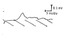 The
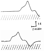
usual motor response has a fairly simple waveform. It
may have one or two initial negative (up) peaks (the
latter usually indicating two muscle being stimulated)
and usually will be followed by a positive deflection
(down) toward the end. The response should have a clear
initial negative deflection as it takes off from the
baseline. In some pathological processes, the wave may
have multiple phases, appearing extremely complex. The
usual motor response has a fairly simple waveform. It
may have one or two initial negative (up) peaks (the
latter usually indicating two muscle being stimulated)
and usually will be followed by a positive deflection
(down) toward the end. The response should have a clear
initial negative deflection as it takes off from the
baseline. In some pathological processes, the wave may
have multiple phases, appearing extremely complex.
The motor response also changes
in relationship to the point of nerve stimulation. The
more proximally the nerve is stimulated, the lower the
amplitude and the longer the duration of responses seen.
These effects are due to the temporal dispersion of
the motor units activated because of differential conduction
velocities in the normal motor nerves.
Sensory-Nerve
Action Potentials
Sensory-nerve action potentials
(NAP) are obtained by stimulating a nerve and recording
directly from it or one of its branches. The recording
site must be remote from muscles innervated by that same
nerve because muscle responses will obscure the much smaller
NAP.
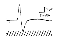 The
NAP can also be characterized by it amplitude, duration,
and wave form. The amplitude of the NAP is measured
from the peak of the positive deflection the peak of
the negative deflection and is measured in microvolts.
The sensory distal latency is traditionally measured
from the stimulus artifact to the takeoff or the peak
of the negative deflection. When conduction velocities
are needed, distal latencies to the takeoff of the proximal
and distal responses should be used. The amplitude depends
on the number of axons being stimulated and the synchrony
with which they transmit their impulse. If the axons
transmit impulses at comparable velocities, the response
duration will be short and amplitude high. However,
if the axonal velocities are widely dispersed, the NAP
duration will be longer and its amplitude lower. The
NAP can also be characterized by it amplitude, duration,
and wave form. The amplitude of the NAP is measured
from the peak of the positive deflection the peak of
the negative deflection and is measured in microvolts.
The sensory distal latency is traditionally measured
from the stimulus artifact to the takeoff or the peak
of the negative deflection. When conduction velocities
are needed, distal latencies to the takeoff of the proximal
and distal responses should be used. The amplitude depends
on the number of axons being stimulated and the synchrony
with which they transmit their impulse. If the axons
transmit impulses at comparable velocities, the response
duration will be short and amplitude high. However,
if the axonal velocities are widely dispersed, the NAP
duration will be longer and its amplitude lower.
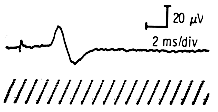
Distal
Latency
Defined as the time from the
stimulus affecting the nerve to the response (motor or
sensory) being recorded, latency is usually measured in
milliseconds (msec). Distal latency is that interval measured
from the stimulation of the distal-most accessible site
on the nerve. This finding does not give direct information
on conduction velocities, because the distal segment often
follows a tortuous route that cannot be measured. The
measurement is useful, however, because it can be compared
with normal data and indicate the relative conductivity
of the segment of the nerve. In measuring the latency
of the motor nerve, remember that a small portion of that
time is due to the delay in neuromuscular transmission,
whereas no such delay is present in sensory latencies.
Conduction
Velocity
If a nerve can be stimulated
at two points along its course, and a measurement can
be obtained of the distance between those points, conduction
velocities can be figured.
 This
is true for most motor nerves. In sensory studies however,
only one stimulation site is nromally used. Compute
the velocity (V) by measuring the distance (d) in millimeters
(mm) between the two stimulation points and dividing
by the difference in latency (ms) between the proximal
(tp) and distal stimulation points (td), as indicated
in this equation: This
is true for most motor nerves. In sensory studies however,
only one stimulation site is nromally used. Compute
the velocity (V) by measuring the distance (d) in millimeters
(mm) between the two stimulation points and dividing
by the difference in latency (ms) between the proximal
(tp) and distal stimulation points (td), as indicated
in this equation:
V=d/tp-td
The result is expressed as meters
per second (m/sec.).
Because the proximal and distal
latencies are measured to the takeoff of the response,
the conduction velocity obtained represents conductions
along the fastest conducting fibers, with those that
first reach the muscle causing the initial deflection.
Conduction velocities in the various
nerves differ, depending on anatomical considerations.
However, several general principles apply to evaluating
nerve conduction studies:
The more proximal the segment
of nerve being evaluated is, the faster the velocity
will be. If the extremity being tested is cold, the
velocity will be slowed and the amplitude increased.
This effect occurs especially in cold weather and some
provisions for warming the patient and for using a fairly
constant room temperature should be made. At times anatomical
considerations such as potential entrapment points will
also tend to slow the velocities. The shorter the segment
between the two stimulation points, the less reliable
the calculated velocities will be, due to a greater
effect on the margin of error by a shorter distance.
Conduction velocities depend most
on the integrity of the myelin sheath. In segmental
demyelinating diseases conduction velocities drop to
below 50 percent of normal values. However, when axonal
loss is severe, the velocity will also be slowed due
to a dropout of the fastest conducting fibers. The drop
in axonal loss is usually in the vicinity of 30 percent
below normal values.
Machine
Settings
In the study of sensory and
motor responses, different filter, sweep speed, and sensitivity
settings are used. Sensory studies are performed with
the low frequency setting between 32 and 50 Hz and the
high frequencies between 1.6-2 and 3 KHz. The sweep speed
is set to 2 ms/division and the sensitivity at 10-20 µV/division.
Motor studies are performed with the low frequency set
to 1.6-2 Hz and the high frequencies to 8-10 KHz. Depending
on the response's latency and duration, the sweep speed
can be set to anywhere between 2-5 ms/division and the
sensitivity between 2-10 mv/division. Whatever the setting,
the distal and proximal latencies should be measured at
the same setting, preferably using the faster sweep speed,
as the takeoff is easier to identify with faster sweeps.
Normal Values
Normal values can be sorted
according to age, sex, extremity length, patient's height
or a combination thereof. Unless otherwise specified,
we use the Cleveland Clinic Foundation's EMG Lab normal
values which were sorted according to patient's age. These
normals were based on a sampling of a minimum of forty
patients for ages ten to nineteen, and seventy and over,
and at least ninety patients for the other age-groups.
The ranges (first two numbers) and averages (between parenthesis)
are provided. These values are based on the following
standard distances: 13 cm for the median sensory (wrist
to active electrode), 11 cm for the ulnar sensory and
10 cm for the radial sensory. For the motor studies, a
minimum of 4-6 cm is used between the wrist and active
electrodes (median and ulnar nerves).
|
|


