No
Response
Sometimes when trying to do
nerve conduction studies you will get no response. Because
such nonresponse can result from many causes, a careful
step-by-step analysis of the nerve stimulation technique
is necessary.
With motor nerve stimulation, you
should see a visible muscle contraction (though in severe
neuropathic disease the contraction may be minimal).
If none is seen follow these steps:
Check to be sure the stimulation
is delivering an impulse. Most patients will feel the
stimulus, but you can check it with your finger while
turning up the voltage. If no stimulus is being delivered,
then check the switches to see if they are on: remove
the stimulator wires from their sockets and reinsert
them properly. Next, check the stimulator wires for
a defect, first visually then electrically with an ohmmeter
to determine whether the wire has continuity. If after
following these steps you find nothing amiss, then the
problem lies within the stimulator, which must be tested
out by the electronics service man.
If the stimulator is found to be working, then check
the anatomical location of the stimulation electrodes.
Occasionally a beginner will place the electrodes in
the wrong area or over the wrong nerve.
If the stimulating electrodes are
in the proper position, then check for the amount of
cream under the anode and cathode. Too much cream or
sweating will create a cathode-anode bridge and will
render nerve stimulation impossible. Try drying the
skin with alcohol or ether. Little or no cream will
deliver a submaximal stimulus strength.
If the stimulating electrodes are
in the proper position, then raise the stimulus strength
to the full output of the stimulator. If there is no
response, increase the duration of the stimulus; next,
bring the stimulus to full strength. This procedure
is often necessary in the extremely obese persons or
in those with edema, severe nerve disease, or regenerating
nerves.
Muscle
Contraction But No Evoked Response
Check the switch controlling
the input on the preamplifer to be sure it is in the "on"
position.
Confirm that the recording electrodes
are over the end-plate area of the muscle being stimulated.
If you still get no response.
Remove excessive cream, which can
cause an active bridge to the reference electrode and
will result in either a very small or no response. Add
cream wherever it is insufficient under the recording
electrode. (Insufficient cream can have the same effect
as too much cream.)
Check the recording electrodes
and connecting wires with an ohmmeter for their integrity
or replace them with new electrodes.
On a multichannel EMG machine,
if you still get no response, check the connections
between the appropriate preamplifer and amplifier.
Check the ground lead, for often
when the ground is not in contact, the trace on the
CRT will be off the screen.
Assure that the trace is centered
on the screen by checking the appropriate channel selection
on the CRT.
Set the adequate CRT sweep speed
so that the expected response is on the screen (Try
using a slower sweep speed to see if the response is
off the screen).
In the event that the response
is of low voltage, increase the gains on the amplifier.
Stimulus
Artifact
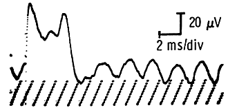
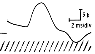
If the record shows a large
stimulus artifact, look into these possibilities:
The ground
is not functioning (sensory potential with loose ground
on the left, motor on the right). Be sure that
the electrode paste is adequate and that the ground
is on tightly and located in the right place, preferably
near but not touching the recording electrodes or between
the stimulating and recording electrodes, and the electrode
wire is tested with an ohmmeter to assure its continuity.
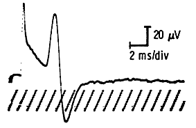
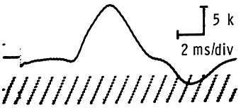
A recording electrode is defective
(Sensory potential with loose active electrode on the
left, motor on the right). Again, be sure the electrode
paste is adequate, the electrodes are on tightly, and
the electrode and wire are checked with an ohmmeter
for a defect. Defective electrodes should be changed.
The electrodes and their wires should also be tested
with an ohmmeter.
Check the stimulating electrodes
to assure that there is no electrode paste bridge between
the electrodes.
If the above measures do not help,
try using needle recording electrodes.
Make sure recording and stimulation
electrode connection cables are not crossed and touching.
Abnormal
Recorded Potential
If the recorded potential is
abnormal in its voltage, follow these steps:
Move
the stimulating electrodes in small increments until
the best response is obtained. Be sure that the stimulus
strength is supramaximal (submaximal stimulus may appear
to give a decremental type of response, especially if
the stimulator is not directly over the nerve).
Check the recording electrodes
to assure they are over the appropriate muscle or nerve
and that the amount of electrode paste is adequate to
avoid a cream bridge effect (see below).
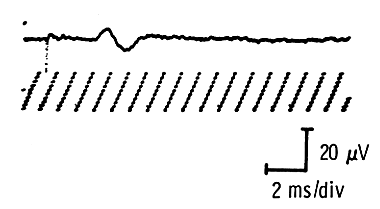
Initial
Positive Deflection
If the evoked response seen
on the cathode ray tube has an initial positive deflection,
do the following, except for the posterior tibial nerve,
where recording from the abductor hallucis (AH) usually
results in an initial positive deflection.
Move
the active recording electrode about until it
is over the motor point of the muscle.
Make sure that the appropriate
nerve is being stimulated and that there is not a spill
over to another, faster conducting nerve (which can
be checked by stimulating that other nerve).
Consider whether a crossover is
present that would stimulate more remote muscles sooner
than the one being tested (see page 59).
Check for reversed electrode
connections to preamplifier input jacks.
|


