The
EMG Manual is a simple step-by-step approach to
the basic understanding of EMG and Nerve Conductions
studies. It is designed to familiarize the reader
with the basic aspects of nerve conduction studies
and needle examinations and can serve as a guide
to the test, a preparation for the EMG rotation,
or as a simple means for better understanding of
the indications, preparations, interpretation and
performance of EMG Nerve and Conduction studies
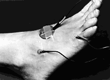
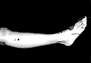
Peroneal NervePlace the recording electrode
over the Extensor Digitorum Brevis (EDB) muscle, over
the lateral aspect of the dorsum of the foot, located
by asking the subject to wiggle their toes. Place the
reference electrode over the base of the little toe.
Stimulate the nerve distally at
the ankle, lateral to the anterior tibial tendon; stimulate
it proximally in the lateral popliteal space.
In the work-up of a peroneal neuropathy,
stimulation below the fibular head should be done to
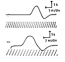
rule out lesions at tat level. When an absent or a low
response is evoked on stimulation of the peroneal nerve
at the ankle (smaller than the response obtained from
proximal stimulation), stimulate the area behind the
lateral malleolus to pick up a variant in the innervation
of the extensor digitorum brevis by way of the accessory
peroneal nerve. If stimulation of either site does not
give a response, then study of the anterior tibial muscle
may give information of peroneal nerve function.
Peroneal
Entrapment, Compression or Injury Sites
The peroneal nerve is most frequently
involved, by pressure or injury, at the fibular head,
causing the classical foot drop; as part of a sciatic
nerve injury; or at the ankle in the anterior tarsal tunnel
(deep branch).