The
EMG Manual is a simple step-by-step approach to
the basic understanding of EMG and Nerve Conductions
studies. It is designed to familiarize the reader
with the basic aspects of nerve conduction studies
and needle examinations and can serve as a guide
to the test, a preparation for the EMG rotation,
or as a simple means for better understanding of
the indications, preparations, interpretation and
performance of EMG Nerve and Conduction studies
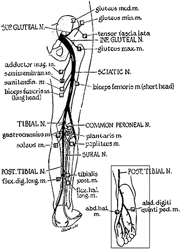
Posterior Tibial Nerve
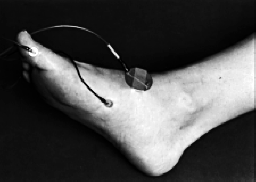
The recording electrode is routinely placed over the abductor
hallucis muscle, located one fingerbreadth behind and
below the navicular bone. You can also place it over the
abductor digiti quinti muscle, the latter placement being
useful for comparing the medial and lateral plantar nerve
functions. Place the reference electrode over the base
of either the great or little toe.
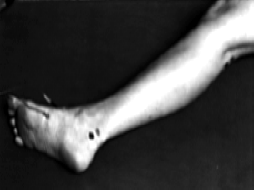
Stimulate the nerve distally posterior
to the medial malleolus at the ankle. The proximal stimulation
point is in the popliteal space, about a fingerbreadth
lateral to midline.
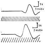
At times, surface stimulation of the nerve in the popliteal
space may be difficult, especially in obese patients.
Often you can get a response if the stimulus voltage
and duration are raised to uncomfortable levels. Often,
too, this response has an initial positive deflection
and its amplitude drops by more than 2 mv. It should
however remain within 4 mv of the distal response. You
may need to position the patient in several ways before
a response is obtained, often the best one being with
the patient prone.
Posterior
Tibial Entrapment, Compression or Injury Sites
The posterior tibial nerve may
be involved as part of a sciatic nerve injury; at the
popliteal fossa; in the tarsal tunnel following ankle
injury; and rarely at an anterior opening of the abductor
hallucis muscle.