The
EMG Manual is a simple step-by-step approach to
the basic understanding of EMG and Nerve Conductions
studies. It is designed to familiarize the reader
with the basic aspects of nerve conduction studies
and needle examinations and can serve as a guide
to the test, a preparation for the EMG rotation,
or as a simple means for better understanding of
the indications, preparations, interpretation and
performance of EMG Nerve and Conduction studies
NERVE CONDUCTION
STUDIES | LOWER EXTREMITY NERVES | THE FEMORAL AND SCIATIC
NERVES
Femoral Nerve
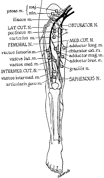
Place the active recording electrode over the vastus medialis
muscle and the reference electrode on the patella.
Stimulate the nerve in the groin
over the femoral triangle or at Hunter's canal. You
can also stimulate it above the inguinal ligament.
Because this nerve is difficult
to stimulate in obese patients, especially above the
inguinal ligament, needle electrodes may be used for
that purpose.
Femoral Entrapment, Compression
or Injury Sites
The posterior tibial nerve may
be involved as part of a sciatic nerve injury; at the
popliteal fossa; in the tarsal tunnel following ankle
injury; and rarely at an anterior opening of the abductor
hallucis muscle.
Sciatic Nerve
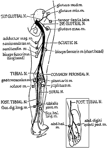
Place the recording electrodes on those muscles used in
peroneal or posterior tibial testing. The stimulating
electrode must be a needle electrode over the sciatic
notch, which is halfway between the ischial tuberosity
and greater trochanter.
Sciatic
Entrapment, Compression or Injury Sites
The sciatic nerve may be injured
high along its course at the roots and plexus level; underneath
the pyriformis muscle; in the buttock (most notoriously
by injury from intramuscular injections); and along its
entire course in the thigh by fracture, missile wounds
orother types of injuries.